Use a point mutation mouse Knockin to circumvent complex phenotypes arising from complete Knockouts (e.g., signaling pathway problems, cross-reactivity).
A point mutation Knockin mouse defines an animal model in which one or more nucleotides are constitutively mutated.
The insertion, deletion, nonsense and sense mutations can alter the amino acid sequence of a given protein, and so dramatically affect its function.
Applications
For academic research:
Study protein function (gain or loss of function)
Analyze the role of non-coding regions and regulatory elements
Investigate disease-causing mutations
For bio-pharmaceutical research & development:
Study drug-resistant mutants
Alter drug-antibody affinities
Pharmacological off-target and efficacy studies
Mimic human genetic diseases
Strengths of point mutation Knockin mouse models
Best way to reproduce human disease when due to mutations
High physiological relevancy of the scientific data obtained from the model (regulatory elements conserved, under control of endogenous promoter, expression of all splice variants, etc.) = cleaner way than classical KO where the whole gene is deleted
Phenotype due only to the mutation: alteration of a single function without disturbing other domains of a protein
Limitations of point mutation Knockin mouse models
Mutation of the gene of interest may affect development, resulting in an impaired phenotype or embryonic death → Limitation can be bypassed by applying conditions such as time-specific gene inactivation
1. Modification or disruption of splicing regulation 2. Genetic redundancy → Can be assessed via constitutive Knockout of the gene of interest
Case Study
Model with heterozygous point mutations, exactly homologous to human retinitis pigmentosa (RP).
Mutations in the PDE6A gene can cause rod photoreceptor degeneration and the blinding disease retinitis pigmentosa.
Model: Novel mouse model for the Pde6aR562W point mutation in combination with an existing line carrying the V685Mpoint mutation to generate compound heterozygous Pde6aV685M/R562W animals, exactly homologous to a case of human RP.
Aim: Predict time-courses for Pde6a-related retinal degeneration and thereby facilitate the definition of a window of opportunity for clinical interventions.
Results: The study provides a rational basis for predictions on human RP phenotypes and disease progression in compound heterozygous situations, and suggests the targeting of non-apoptotic processes as a feasible treatment approach.
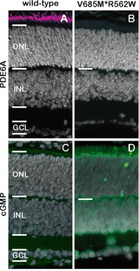
Figure 1. Loss of PDE6A expression causes cGMP accumulation.
Excessive accumulation of cGMP and subsequent rod photoreceptor death, followed by a mutation-independent, secondary death of cone photoreceptors.
A, B) In the PN11 wildtype (wt) retina (A), immunostaining for PDE6A shows strong protein expression in photoreceptor OS.
In contrast, at the same age, in the compound heterozygous V685M/R562W retina (C), the protein is essentially absent.
C, D) At PN11, wt retina is essentially negative for cGMP immunoreactivity (C).
PDE6A mutants, however, display individual rod photoreceptor cells that have accumulated large amounts of cGMP (D).
E) The quantification of cGMP-positive cells in the ONL and the PDE6A pixel intensity in the OS (arbitrary units; AU) shows an inverse correlation. Images are representative for immunostaining performed on retinal sections from at least three independent animals for each genotype.
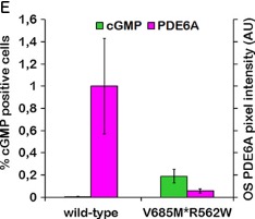
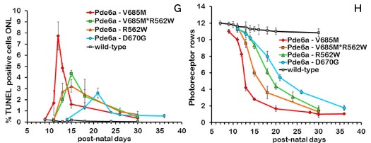
Figure 2. Photoreceptor cell death and survival.
A-C) The TUNEL assay in the wt retina occasionally labeled cells dying due to developmental processes.
D-F) In Pde6a mutants, photoreceptor cell death was dramatically increased.
The images show the situation at P12, P15 and P21, time-points corresponding to the peak of cell death..
G, H) The line graph at the bottom left (G) illustrates the progression of photoreceptor cell death as evidenced by the TUNEL assay in different Pde6a mutants. The green line corresponds to the V685M*R562W mutant.
The peak times as well as the peak amplitudes correspond to the speed of retinal degeneration, which is illustrated by the loss of photoreceptors (H).
Images are representative for TUNEL assays performed on retinal sections from at least three independent animals; quantifications in G, H include data from 3-7 animals per genotype and time-point.
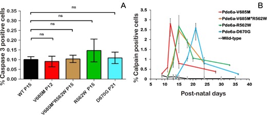
Figure 3. Photoreceptor degeneration in Pde6a mutants correlates with calpain, not caspase, activity.
Calpain activity is often associated with non-apoptotic forms of cell death, and in all Pde6a mutants it is strongly correlated in time with the progression of photoreceptor cell death.
A) Quantification of caspase-3-positive cells, which shows only extremely low numbers of cells at the respective peaks of degeneration, with no significant (n.s.) differences to wt.
B) The progression of calpain activity was analyzed over time and showed a strong correlation to the extent of cell death and the progression of retinal degeneration.
Oops! Something went wrong while submitting the form.
Mouse
Knockin
Permissive locus
Customized
Quick KI mouse
The Rosa26 and Hprt gene loci are well suited for gene over-expression, reduced development time and cost with ready-to-use targeting vectors.
Mouse
Knockin
Reporter
Customized
Reporter KI mouse
Use a reporter mouse Knockin for in vivo monitoring of transcriptional promoter activity, protein localization, cell trafficking, etc.
Mouse
Knockin
Customized
Humanized KI mouse
Use humanized mice as in vivo tools for mimicking human pathological conditions and diseases, and for conducting preclinical research.
Mouse
Knockout
Point mutation
Customized
Protein function KO mouse
A protein function Knockout mouse defines a model in which one or more nucleotides are mutated in a way that the protein loses its function.
Mouse
Knockout
Constitutive
Customized
Constitutive KO mouse
A constitutive, conventional, or whole-body Knockout mouse is a fast and cost-effective solution for in vivo preliminary studies of target gene functions.
Mouse
Knockout
Conditional
Time-dependent
Customized
Time-dependent KO mouse
Use an inducible conditional Knockout mouse to age-dependently inactivate your gene, and to enable studies at defined development stages or on age-related diseases.
Mouse
Knockout
Conditional
Tissue-specific
Customized
Tissue-specific KO mouse
Use tissue- or cell-specific conditional Knockout mouse models to bypass embryonic lethality, compensatory mechanisms, complex phenotypes, etc.
Oops! Something went wrong while submitting the form.
Ultimate predictive models
Physiological relevance Comprehensive and reliable validation data Over 600 scientific articles based on our models
Scientific excellence
In-depth expertise in preclinical science, physiology, and model development Unique R&D platform located in France
Customer-centric
Tailor-made approach to your needs Strong emphasis on customer satisfaction
Collaborative partner
Long-standing partnerships with 17 of the top 20 pharma companies Co-development of innovative models for next-generation drugs Guaranteed FTO/freedom to operate
.png)
.png)