.png)


Applications of the genO‑hPD‑1/hLAG3 mouse model
Useful applications for researchers working in immuno-oncology and infectious diseases:
- Assess the efficacy of PD-1 and/or LAG3-targeting agents, including tumor growth inhibition studies and the discovery of new therapeutic approaches
- Understand the mechanism of action (MoA) of different constructs and their possible therapeutic advantages
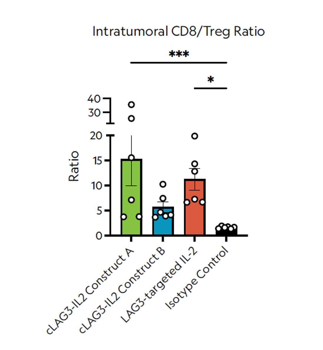
- Develop antibody based therapies aimed at targeting the tumor microenvironment, as hLAG3 is observed in the TME of genO‑hPD‑1/hLAG3 mice
Features
- hPD-1 and hLAG3 expression driven by the respective endogenous mouse promoter
- Fully functional mouse immune system
- Lack of expression of the murine target gene, thus avoiding cross-reactivity
Clients