.avif)


Applications of the genO‑hIgE/hFcεR1 mouse model
Useful applications for researchers working in immuno-inflammation and allergic diseases:
- Assess the efficacy of IgE-targeting therapeutics
- Test therapeutics that block anaphylaxis (passive or active) and help controling asthma
- Develop models of Passive Cutaneous Anaphylaxis (PCA), OVA hypersensitivity and Passive systemic anaphylaxis (PSA)
- Investigate the mode of action (MoA) of various compounds and understand their therapeutic potential
Features
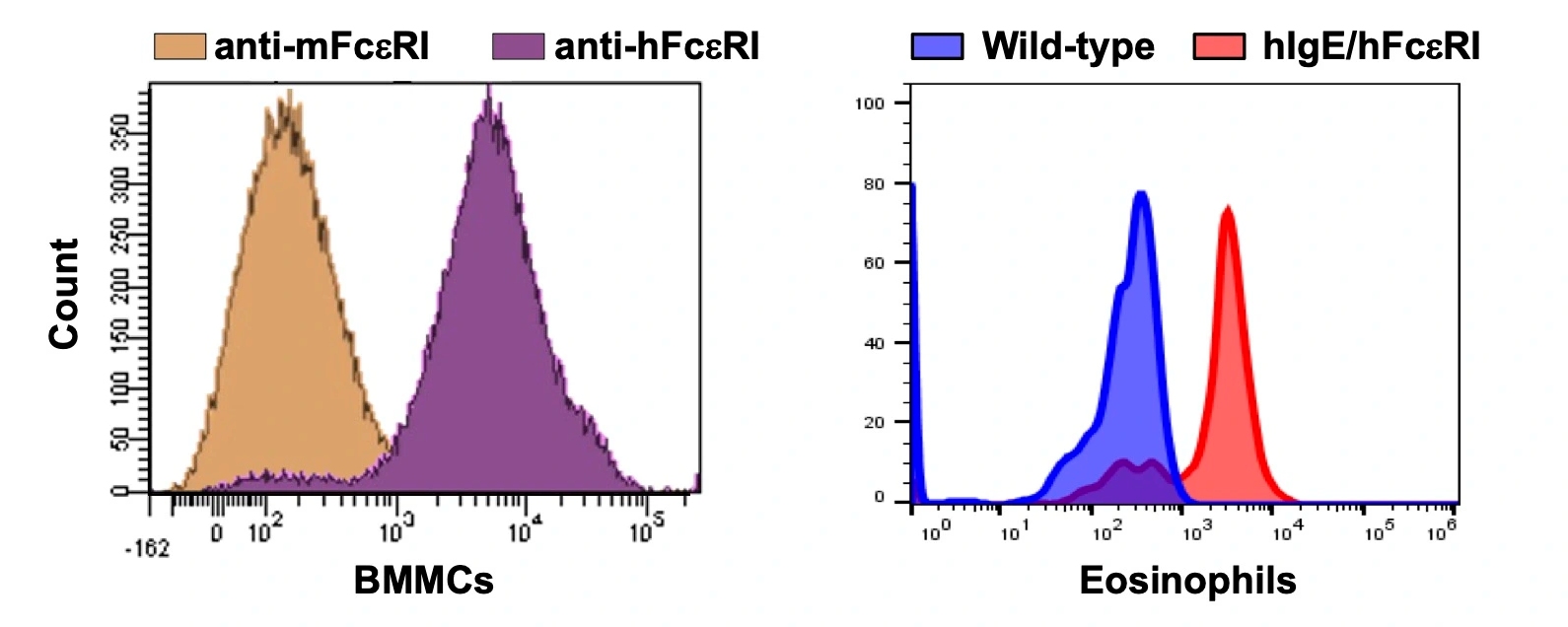
- Human-like cellular distribution (i.e., mast cells, basophils, monocytes/DCs, Langerhans cells, and eosinophils) of the FcεR1 receptor
- Physiological regulation and expression of the chimeric IgE/FcεR1 complex
- Preservation of ligand-receptor interaction
- Preservation of the Ig class switching mechanism
- Human IgE is present, but at low levels
- Fully functional mouse immune system
- Lack of expression of the murine target genes
Clients