Related resources and publications

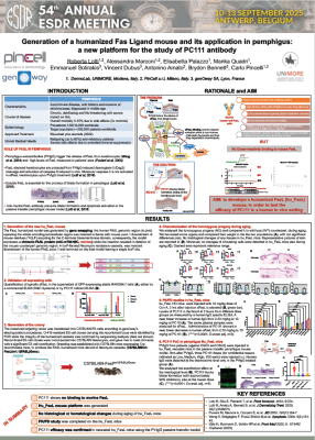
Antibody efficacy in customized hFasL mouse

CD28xVISTA bispecific antibodies in immunotherapy

Safety and activity of a conditionally active cLAG3-IL2

Multispecific CD73/PD-1 targeting

FcγR mouse models for efficacy and PK/PD

CD28-targeting therapies

Drug delivery to the brain (TFRC)

BRGSF-HIS & hFcγR to assess cell depletion

CD3 binding T-cell engagement

Novel PD-1 targeted IL-2Rβ/γ agonist

Humanized Mouse Model of Fcγ Receptors

hPD-1 to evaluate novel cancer therapies

PD-1 & strong tumor growth inhibition

CTLA-4 & survival in a colorectal cancer model

Anti hCD39 to promote anti-tumor immunity

CD3 and BRGSF-HIS to assess IrAEs

BRGSF-HIS & hVISTA for immunotherapies

VISTA in the tumor microenvironment

hSTING & anti-tumor response in cold tumors

BRGSF-HIS & CD3ε in CRS

anti-hVISTA against poorly immunogenic tumors

Sci Immunol
Cancer Immunol Res
J Immunother Cancer
J Nucl Med
Microbiol Spectr
J Immunother Cancer
Sci Adv
Cancer Immunol Res
Mol Cancer Ther
Nat Commun
J Clin Invest
Alzheimers Res Ther
Cancer Immunol Res
Theranostics
Eur J Nucl Med Mol Imaging
JCI Insight
JCI Insight
Mol Cancer Ther
Clin Cancer Res
Nat Commun
Hum Mol Genet
Front Immunol
Front Immunol
Hear Res
Front Immunol
Cell Mol Life Sci
Front Immunol
J Allergy Clin Immunol
Proc Natl Acad Sci U S A
Immunohorizons
PLoS One
J Thromb Haemost
Pancreatology
Acta Biomater
Matrix Biol
Front Immunol
J Invest Dermatol
J Allergy Clin Immunol
Int J Pharmac
J Immunother Cancer
Prog Neurobiol
Nat Cancer
Cell Rep
Sci Rep
Mol Cancer
Neurobiol Dis
ACS Pharmacol Transl Sci
Mol Cancer Ther
Cell Death Dis
Nat Med
Matrix Biol
Bioconjug Chem
Proc Natl Acad Sci U S A
Cancer Discov
J Bone Miner Res
Nat Chem Biol
Cell
Nat Chem Biol
J Immunother Cancer
Sci Adv
Hum Genet
Eur J Microbiol Immunol
J Alzheimers Dis
Cell Physiol Biochem
Invest Ophthalmol Vis Sci
JCI Insight
PLoS One
EMBO J
Circ Res
Brain
PLoS One
J Biol Chem
J Am Heart Assoc
J Control Release
Arterioscler Thromb Vasc Biol
Physiol Rep
Blood
J Bone Miner Res
J Neurosci
Cell Death Differ
Cell Mol Life Sci
Diabetes
Humanized ICP in immuno-oncology
Immuno-oncolology models
Immuno-oncolology models
Humanized mouse models
T-cell engager
Assess toxicity with ICP
Bispecific antibodies
Physiological relevancy in preclinical models
Therapeutic efficacy and pharmacokinetics
T-cell engager
Humanized PD-1 & GITR mouse
T-cell engager CD3
Assess T-cell engager in BRGSF-HIS
Humanized PD-1/PD-L1 mouse and cell line
Humanized HSA/hFcRn mouse
Humanized ICP mouse and cell lines
Assess T-cell engager in BRGSF-HIS
Humanized VISTA mouse & Pharmacokinetics
Humanized cLC model - Bispecific antibody development
Targeting the human CD47/SIRPα axis
BRGSF-HIS as model for acute implant-associated osteomyelitis
VISTA - safety and PK profiling
CD3ε - T-cell engager assessment
TFRC for anti-cancer therapies
CD4 for cancer treatment outcomes
hVISTA - antibodies & PET imaging


